techniques: Electron Microscopy
The cornerstone of synthetic chemistry and biochemistry research, these tools allow the determination of chemical structure and molecular environment. Truly state of the art, SHyNE’s molecular characterization tools include NMR, mass spec, chromatography and more.
SCANNING ELECTRON MICROSCOPY (SEM)
 Scanning electron microscopy (SEM) uses a focused electron probe to scan the surface of a sample. The electrons interact with the surface of the sample to produce a wide range of useful signals that can be used to provide high spatial resolution images, elemental composition, crystallographic information and more. Our instruments are capable of analyzing more traditional hard materials (metals, ceramics, etc.) as well as non-conductive or even hydrated samples by variable pressure/environmental SEM.
Our current range of SEM instruments can be found in our EPIC Facility.
TRANSMISSION ELECTRON MICROSCOPY (TEM)
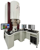 Transmission electron microscopy (TEM) uses a high energy electron beam which is transmitted through a thin sample to provide information via the interaction of electrons with your sample. Modern TEM’s are a platform for a range of different types of experiments from atomic resolution imaging to structural analysis by electron diffraction to in situ or in operando methods, which provide controlled environments and allow application of stimuli to materials inside the microscope. Beyond imaging, TEM can be operated in Scanning TEM (S/TEM) mode to provide high spatial resolution elemental, chemical information and crystallographic information.
Our current range of TEM instruments can be found in our EPIC-TEM Facility.
CRYO-ELECTRON MICROSCOPY
For hydrated materials or beam-sensitive materials, cryogenic methods can be used to preserve structure and provide critical morphological or compositional information. Our facilities house a complete array of cryogenic sample preparation equipment and capabilities for cryo-TEM and cryo-SEM.
Our current range of Cryo-EM instruments can be found in our BioCryo Facility.
ELECTRON MICROANALYSIS
The bevvy of signals produced by the electron-specimen interaction can provide a range of useful information about your sample. X-ray microanalysis techniques such as energy or wavelength dispersive spectrometry (EDS or WDS) are available on many of our systems to provide high spatial resolution elemental analysis. In addition, some of our TEM systems are equipped with electron energy loss spectrometers (EELS) for elemental and chemical analysis. We also have crystal orientation mapping capabilities by electron backscatter diffraction (EBSD).
Our current range of microanalysis instruments can be found in both our EPIC-SEM Facility and our EPIC-TEM Facility.


Dr. Reiner Bleher
BioCryo Facility Manager, Assistant Research Professor
Office: Hogan Hall 5-156
(847) 467-3540 / Email


Dr. Roberto dos Reis
Research Assistant Professor, Scientific Officer Dravid Group
Office: Tech AG90
(847) 467-0105 / Email